Target Gene Search For Mir
Genome-wide screens using miR-874 transfectants were performed to identify target genes of miR-874 in IMC-3. Oligo-microarray human 44K was used for expression profiling of the transfectants in comparison with a miRNA-negative control transfectant. Hybridisation and wash steps were performed as previously described . The arrays were scanned using a Packard GSI Lumonics Scan Array 4000 . The data were analysed by means of DNASIS array software , which converted the signal intensity for each spot into text format. The log2 ratios of the median subtracted background intensities were analysed. Data from each microarray study were normalised by a global normalisation method. Predicted target genes and their target miRNA binding site seed regions were investigated using TargetScan . The sequences of the predicted mature miRNAs were confirmed using miRBase .
Blood Supply And Lymphatics
Paranasal Sinuses
The anterior and posterior ethmoidal branches of the ophthalmic artery supply the ethmoidal and frontal sinuses. The infraorbital artery and the superior, anterior, and posterior alveolar branches of the maxillary artery supply the mucosa of the maxillary sinus. The pharyngeal branch of the maxillary artery supplies the sphenoidal sinus.
Management Of Malignant Tumors Of The Sinuses
As with other types of cancers, a multimodality approach in consultation with a tumor board is recommended in sinonasal malignancies , including a head and neck surgeon and a neurosurgeon when indicated and a neuroradiologist, pathologist, radiation oncologist, and medical oncologist as active members.
Operative approaches to sinonasal malignancies include the following:
- Medial maxillectomy endoscopic
Treatment for recurrence includes surgery, chemotherapy, and/or radiation therapy.
Also Check: Best Way To Treat Sinus Pressure
American Joint Committee On Cancer Stage Groupings And Tnm Definitions
Staging for nasal cavity and paranasal sinus carcinomas is not as wellestablished as staging for other head and neck tumors. For cancer of the maxillary sinus, the nasal cavity, and the ethmoid sinus, theAJCC has designated staging by TNM classification. Lymphomas, sarcomas, and mucosal melanomas of the paranasal sinuses and nasal cavity are not staged using this system. The staging described below is used only for patients who have not had a lymph node dissection of the neck.
| M Category | M Criteria |
|---|---|
| aReprinted with permission from AJCC: Nasal cavity and paranasal sinuses. In: Amin MB, Edge SB, Greene FL, et al., eds.: AJCC Cancer Staging Manual. 8th ed. New York, NY: Springer, 2017, pp 13747. | |
| M0 | No distant metastasis . |
| M1 |
| Stage | Description |
|---|---|
| T = primary tumor N = regional lymph node M = distant metastasis. | |
| aReprinted with permission from AJCC: Nasal cavity and paranasal sinuses. In: Amin MB, Edge SB, Greene FL, et al., eds.: AJCC Cancer Staging Manual. 8th ed. New York, NY: Springer, 2017, pp 13747. | |
| 0 | |
| N0 = No regional lymph node metastasis. | |
| M0 = No distant metastasis . |
References
Small Cell Neuroendocrine Carcinoma

Small cell neuroendocrine carcinoma , similar to oat-cell carcinoma of the lungs, is reported to arise in the nasal cavity and paranasal sinuses in patients ranging aged 26-77 years. The fact that the tumor is almost always in an advanced stage by the time it comes to attention reflects it aggressive nature. Several sinuses are nearly always involved. Cervical lymph nodes and pulmonary metastases may also be involved.
Treatment is multimodal, including regiment combinations of surgery, chemotherapy, and radiation therapy. Despite maximal efforts and individualized therapy, the prognosis is poor. Median survival, as extrapolated from several studies, is less than 2 years.
Don’t Miss: South Florida Sinus And Allergy Center
Treatment Outcomes And Related Toxicities
The overall 5-year survival rate for patients with maxillary sinus cancer is 30% to 50%. Cervical metastases occur in less than 10% of maxillary sinus cancer thus a prophylactic neck dissection is not indicated in the N0 neck. When present, cervical metastasis is associated with a very poor prognosis with a 5-year survival rate of less than 10%.
Given the complex anatomy and confined location of paranasal sinus malignancies, it is not surprising that treatment-related morbidity is high. Mendenhall and colleagues218 reported that 26% of patients who underwent surgery followed by PORT experienced severe complications, including ipsilateral blindness , postoperative infection necessitating hospitalization , graft failure, frontal bone osteoradionecrosis, frontal lobe necrosis, intracranial bleeding, postoperative meningitis, cerebrospinal fluid leak, nasal bone necrosis, bilateral blindness, and fatal infected bone flap. Chen and colleagues219 demonstrated that over time the observed incidence of grade 3 or 4 late-occurring toxicity decreased from 53% to 45% to 39% to 28% to 16% among patients treated in the 1960s, 1970s, 1980s, 1990s, and 2000s, respectively. They concluded that improvements in the therapeutic ratio were responsible for the decreasing incidence of compilations treated throughout these decades.
Sapna Lohiya, … Jasjit K. Dillon, in, 2018
Mature Mirna Transfection And Small
The following RNA species were used in this study: mature miRNAs, pre-miR miRNA precursors , negative control miRNA , small-interfering RNA , and negative control siRNA . RNAs were incubated with Opti-MEM and Lipofectamine RNAiMax reagent as described previously . Transfection efficiency of pre-miR in cell lines was confirmed based on downregulation of TWF1 mRNA following transfection with miR-1 as previously reported .
Recommended Reading: Children’s Sinus And Cough Medicine
Clinical Evaluation And Follow
Pretreatment evaluation and staging, as well as the need formultidisciplinary planning of treatment, is very important. Generally, thefirst opportunity to treat patients with head and neck cancers is the mosteffective, although salvage surgery or salvage radiation therapy,as appropriate, may occasionally be successful.
Because most treatment failures occurwithin 2 years, patients must be monitored frequently and meticulouslyduring this period. In addition, because nearly 33% of these patientsdevelop second primary cancers in the aerodigestive tract, lifetime follow-up is essential.
Squamous Cell Carcinoma Of Maxillary Sinus: A Comparison Of Clinico
C. P. Sudheer, T. Dinesh Singh
Abstract
Background: The nasal cavity and paranasal sinuses are the site of origin of more complex, histologically diverse group of tumors which include neoplasms derived from mucosal epithelium, seromucinous glands and haematolymphoid cells. This study aimed to assess the incidence and prognosis of maxillary sinus malignancy.
Methods: A total 25 malignancy cases between age group 11-70 years. All the cases were undergone with detailed clinical examination. The TNM staging system was followed to detect and standardize the disease stage. CT scan was used to identify the growth and extent of tumor.
Results: Majority cases were between the age group 41-50 years . Epistaxis was the commonest clinical symptom in 72% cases followed by nasal obstruction , oral related symptoms , oedema in cheeks and auditory disturbances in 16% cases. The TNM Staging system showed that 76% cases had T3 lesions, 16% had T4 lesions and 8% had T2 lesions. Nasal symptoms are commonly associated with malignancies and were belongs to T3 stage and few under T4.
Maxillary sinus malignancy is an aggressive tumor normally diagnosed at the advanced stage, with minimal prognosis and reduced survival rate. Surgery in combination with radiotherapy followed by chemotherapy remains the best treatment modality.
Keywords
Full Text:
References
Grant RN, Silverberg E. Cancer Statistics 1970. American Cancer Society, New York, 1970.
Don’t Miss: Over The Counter Sinus Infection Relief
Squamous Cell Carcinoma Of The Nasal Cavity And Paranasal Sinuses
Disease definition
A rare head and neck tumor characterized by a malignant epithelial neoplasm most commonly arising in the maxillary sinus or nasal cavity, occurring as a keratinizing, a non-keratinizing, or a spindle cell type. Patients may present with nasal obstruction, epistaxis, rhinorrhea, swelling, or with facial pain and/or paralysis, diplopia, and proptosis. Patients with paranasal sinus tumors present later and at a higher stage than patients with nasal cavity carcinomas. Risk factors are smoking and industrial exposures. High-risk HPV is most frequently associated with the non-keratinizing type.
Purpose Of This Summary
This PDQ cancer information summary for health professionals provides comprehensive, peer-reviewed, evidence-based information about the treatment of adult paranasal sinus and nasal cavity cancer. It is intended as a resource to inform and assist clinicians in the care of their patients. It does not provide formal guidelines or recommendations for making health care decisions.
Recommended Reading: Sinus Pressure And Ear Pain
Plasmid Construction And Dual
The wild-type sequences of PPP1CA 3-UTR and those with deleted miR-874 target sites were inserted between the XhoIPmeI restriction sites in the 3-UTR of the hRluc gene in psiCHECK-2 vector . Sequences of oligonucleotides are described in the . The synthesised DNA was cloned into the psiCHECK-2 vector. The IMC-3 cells were transfected with 15ng of vector, 10nM of miR-874 , and 1l of Lipofectamine 2000 in 100l of Opti-MEM . The activities of firefly and Renilla luciferases in cell lysates were determined with a dual-luciferase assay system . Normalised data were calculated as the quotient of Renilla/firefly luciferase activities.
Stage Information For Paranasal Sinus And Nasal Cavity Cancer

In This Section
The staging systems are clinical estimates of the extent of disease. Theassessment of the tumor is based on inspection, palpation, and direct endoscopywhen necessary. The tumor must be confirmed histologically, and any otherpathological data obtained on biopsy may be included. The appropriate nodaldrainage areas are examined by careful palpation. Computed tomographic and/ormagnetic resonance imaging studies are generally required to adequatelyevaluate tumor extent before surgical resection or definitiveradiation therapy is attempted. If a patient relapses, complete restaging must be done to select the appropriate additional therapy.
Recommended Reading: Best Medicine For Sinus Congestion And Cough
Recurrent Paranasal Sinus And Nasal Cavity Cancer
In This Section
Chemotherapy for recurrent head and neck squamous cell cancer has shownpromise. Chemotherapy may be indicated when there is recurrence in eitherdistant or local disease after primary surgery or radiation therapy, and when there isresidual disease after primary treatment. Survival may be improved inthose achieving a complete response to chemotherapy. Combined-modalitytherapy with platinum and radiation therapy has been used in clinical trials such as UMCC-8810.
Toxicity And Statistical Analysis
Toxicities were graded using the toxicity criteria of the Radiation Therapy Oncology Group and the European Organization for Research and Treatment of Cancer . Overall survival , local control , regional control and distant control rates were calculated using the KaplanMeier method. Data for patients who were alive or dead without each type of recurrence were used as censored data in calculating LC/RC/DC rates. In calculating local recurrence-free survival rate, local recurrence and any type of death were defined as an event. Parameters such as age, sex, performance, T classification, BED, technique and use of chemotherapy were evaluated to identify prognostic factors using log-rank statistics and Cox’s proportional hazard models.
Read Also: Home Remedies To Get Rid Of Sinus Congestion
Regression Of Advanced Maxillary Sinus Cancer With Orbital Invasion By Combined Chemotherapy And Immunotherapy: A Oneyear Followup Case Report
This article is mentioned in:
Abstract
Introduction
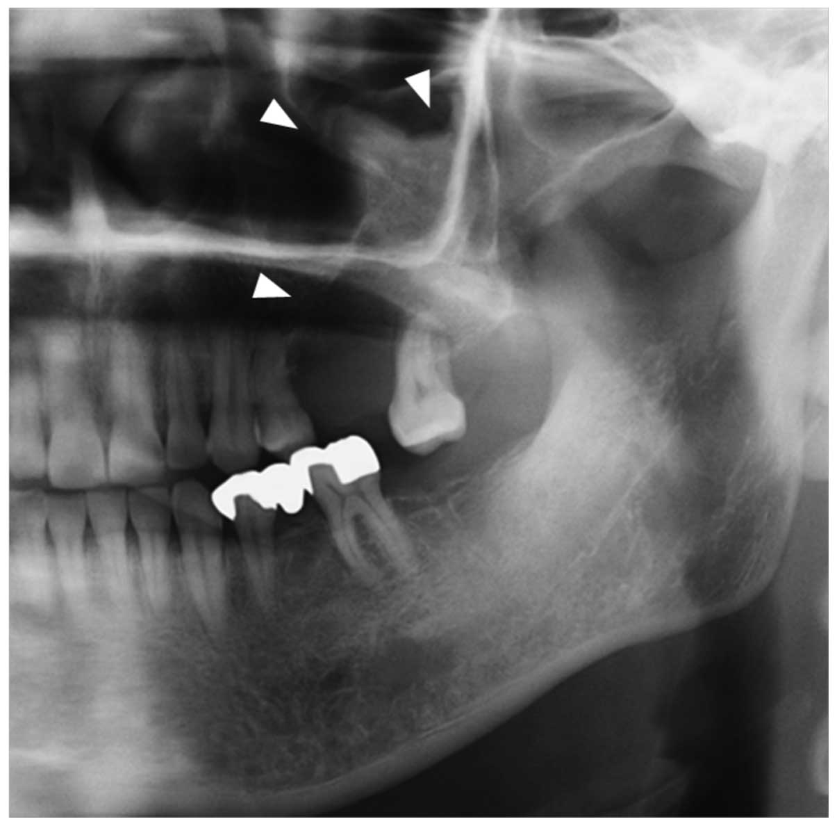
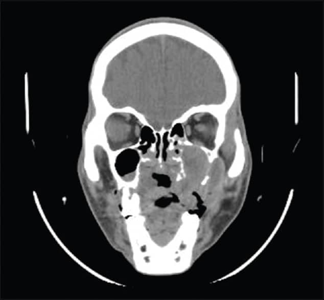
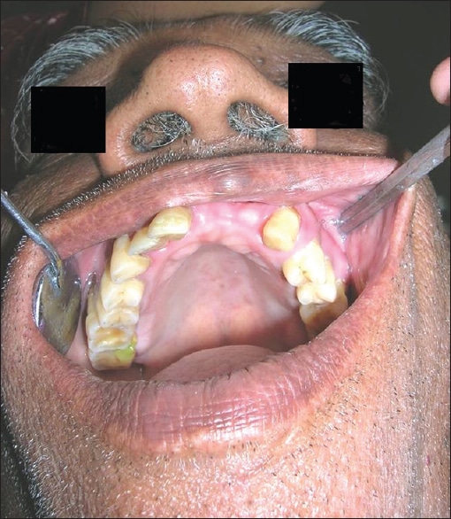
Malignant tumors of the maxillary sinus are rareneoplasms that account for ~3% of head and neck cancers and 0.5% ofall malignant diseases. The annual incidence of maxillary sinuscancer is 0.5-1 case per 100,000 of the population . Squamous cell carcinoma is the mostcommon histologic type, accounting for ~70-80% of maxillary sinuscancer . Most patients withmaxillary sinus cancer have no symptoms in the early stage and,therefore, many of these patients are diagnosed in the advancedstage of the disease . At thetime of diagnosis, 70-80% of maxillary sinus carcinomas presentwith local extension and are classified as T3 or T4 using theAmerican Joint Committee on Cancer staging system .
Maxillary sinus squamous cell carcinoma is anaggressive tumor and treatment includes surgery, radiation therapy,or a combination of both. Nevertheless, for more advanced stages,both surgery and radiotherapy have their limitations . Locally advanced carcinoma of themaxillary sinus has been reported to have a poor prognosis. Induction chemotherapy may beeffective for locally advanced and technically unresectablemaxillary sinus cancers resulting in successful surgery in asignificant proportion of patients .
Case report
Pain In The Nonsurgical Treatment Of Head And Neck Cancer
Pain as an early presenting symptom in head and neck cancer depends very much on the site of the primary tumor. An ulcerated cancer on the lateral edge of the tongue will develop pain early in the disease course, but a maxillary sinus cancer may grow to considerable size before causing any discomfort. Pain can result from direct nerve infiltration by the primary tumor or from the mucositis seen with either chemotherapy or radiotherapy. Pain from the treatment-induced mucositis usually progresses with the development of mucositis minimal erythematous change in the oral mucosa should not give severe pain, and swabs should be taken to exclude fungal super infection. Pain can also indicate bony or dental involvement by tumor, as well as soft tissue bacterial or fungal infection. Following radiotherapy or chemoradiotherapy, pain may be the first symptom of recurrence and should be investigated if presenting at follow-up, especially if it is an unexplained pain or pain presenting in an unusual manner in a patient who has previously been pain free. In this clinical situation, tumor recurrence, potential osteoradionecrosis, or dental pathology secondary to treatment needs to be considered.
Allen M. Chen MD, … A. Dimitrios Colevas MD, in, 2010
Recommended Reading: Will My Sinus Infection Go Away On Its Own
Patients And Tumor Characteristics
Forty-four patients with locally advanced , non-metastatic SCC of MS and NC were treated with definitive radiotherapy at Seoul National University Hospital from May 1990 to May 2007. Although surgical resection followed by radiotherapy with or without chemotherapy is the treatment policy, patients with unresectable or medically inoperable tumors or patients reluctant to undergo surgery were treated with definitive radiotherapy. Excluding two patients who underwent incomplete radiotherapy, we retrospectively reviewed records of 42 patients. The median follow-up time for living patients was 38 months . Clinical tumor staging was evaluated by computed tomography and/or magnetic resonance imaging . Most tumors invaded multiple adjacent structures, and the sites of tumor origin could not be clearly defined in some cases. The site of origin was assigned to the location of the epicenter of a tumor based on CT or MRI findings in these cases. Thirty patients had an MS cancer and 12 patients had an NC cancer. All tumors were histologically confirmed as SCC. Patient and tumor characteristics are listed in Table .
Cellular Classification Of Paranasal Sinus And Nasal Cavity Cancer
The most common cell type for paranasal sinus and nasal cavity cancers issquamous cell carcinoma. Minor salivary gland tumors comprise 10% to 15% ofthese neoplasms. Malignant melanoma presents in less than 1% of neoplasms inthis region. Some 5% of cases are malignant lymphomas.
Esthesioneuroepithelioma, sometimes confused with undifferentiated carcinoma orundifferentiated lymphoma, arises from the olfactory nerves.
Chondrosarcoma, osteosarcoma, Ewing sarcoma, and most soft tissue sarcomashave been reported for this region.
Inverting papilloma is considered a low-grade benign tumor with a tendency torecur and, in a small percentage of cases, to transform into a malignant tumor.
Midline granuloma, a progressively destructive condition, also involves this region.
References
Also Check: Advil Cold And Sinus Rite Aid
Microrna Expression Signatures And Data Normalisation
Tissue specimens for miRNA screening using a low density array were from five MSSCC patients at Chiba University Hospital between 2005 and 2007 . The miRNA expression patterns were evaluated using the TaqMan LDA Human microRNA Panel v2.0 . The assay was composed of two steps: generation of cDNA by reverse transcription and a TaqMan real-time PCR assay. Description of real-time PCR and the list of human miRNAs can be found on the company’s website . Analysis of relative miRNA expression data was performed using GeneSpring GX version 7.3.1 software according to the manufacturer’s instructions. A cutoff P-value of < 0.05 was used to narrow down the candidates after global normalisation of the raw data. After global normalisation, additional normalisation was carried out with RNU48.
Lymphomas And Related Conditions

This category of malignant neoplasia of the sinuses and nasal cavity is complicated, poorly understood, evolving, controversial, and extensive.
In general, non-Hodgkin lymphomas are primarily found in patients in their 60s and 70s and manifest with symptoms of obstruction. Rhinorrhea and epistaxis may also be present. After the type of tumor is established, treatment is usually radiation therapy and chemotherapy, as established by protocol. The prognosis in general is variable for patients with non-Hodgkin lymphoma and, depending on the type and stage ranges, median survival ranges from less than 1 year to close to 80% at 5 years.
Another controversial type of malignant lymphoid tumor is T-cell/natural killercell lymphoma. It has had numerous names throughout its history, including lethal midline granuloma, midline malignant reticulosis, lymphomatoid granulomatosis, angiocentric lymphoproliferative lesion, and T-cell/natural killercell lymphoma. Given the current knowledge, this lesion is probably best categorized as a T-cell/natural killercell lymphoma.
The tumor is a destructive sinonasal lesion associated with obstructive symptoms, bone and soft-tissue destruction, and hemorrhage. It is strongly associated with the Epstein-Barr virus and is most common in Asia and Latin America, with a patient age at presentation of 13-80 years.
Also Check: How To Naturally Fight Sinus Infection