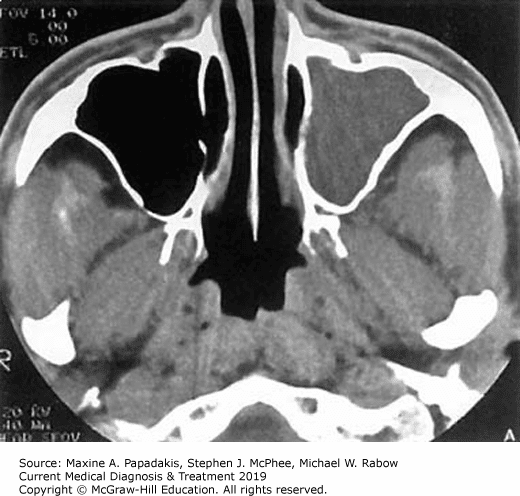
Malignant Disease Of The Maxillary Sinus
Malignancy arising within the paranasal sinuses is relatively rare, constituting 1.0% of all malignancies, with approximately 80% of these malignancies arising in the maxillary sinus with a lesser prevalence in the ethmoid sinus. Malignant disease of the sphenoid and frontal sinuses is very rare. Almost 80% of malignancies are squamous cell carcinomas, with acinic cell carcinomas causing 10%. Metastatic disease presents in the bone and expands into the sinus space.
Malignant disease of the paranasal sinuses unfortunately often presents at a late stage when the tumour has become large enough to cause symptoms. The mucosa of the paranasal sinuses is not as easily accessible as the oral mucosa for routine inspection and early mucosal abnormalities are not seen or investigated.
The dental professional can play a role in the diagnosis of a patient with maxillary sinus malignancy. A combination of patient symptoms and clinical signs should arouse suspicion of maxillary sinus malignancy, warranting immediate referral to an appropriate specialist. Unfortunately, it has been known for patients to be treated for long periods on the assumption that their symptoms arise from chronic inflammatory rhinosinal disease, only for a diagnosis of malignancy to be made at a later date.
Table 4 Some signs and symptoms that may be suspicions for maxillary sinus malignancy
Is Opacification Of Sinus Bad
What causes complete opacification of maxillary sinus?
Mucoceles are most often caused by sinus ostial obstruction that leads to complete opacification of the sinus because of an accumulation of secretions. In many instances, the entire sinus may be expanded on radiologic imaging.
Is ethmoid sinusitis dangerous?
If sinusitis symptoms go on for more than a few days, a doctor will likely prescribe antibiotics to help the infection clear up more quickly. In rare instances, people with numerous infections associated with sinusitis may need surgery to correct any abnormalities. Ethmoid sinusitis complications are rare.
What causes opacification?
The opacification is caused by fluid or solid material within the airways that causes a difference in the relative attenuation of the lung: transudate, e.g. pulmonary edema secondary to heart failure. pus, e.g. bacterial pneumonia. blood, e.g. pulmonary hemorrhage.
What Is Total Opacification Of The Maxillary Sinus
Fact Checked
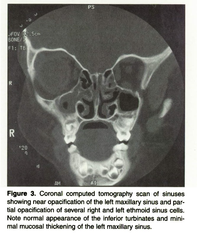
The maxillary sinus is the cavity behind your cheeks, very close to your nose 1.When a CT scan is taken of the head, the sinuses should show up black since they are cavities. When the area shows up white or gray, it is called opaque or opacification of the sinus.
If you are experiencing serious medical symptoms, seek emergency treatment immediately.
Recommended Reading: Severe Sinus And Cold Relief
What Does Near Complete Opacification Of Sphenoid Sinus Mean
Ask U.S. doctors your own question and get educational, text answers â it’s anonymous and free!
Ask U.S. doctors your own question and get educational, text answers â it’s anonymous and free!
HealthTap doctors are based in the U.S., board certified, and available by text or video.
Is Sinus Opacification Serious

Isolated sphenoid sinus opacifications are clinically important because they can lead to serious complications. However, some patients with ISSOs are asymptomatic, and not all patients are properly referred to the otolaryngology department.
How do you get maxillary sinusitis?
The maxillary antrum It is a bacterial infection and typically occurs after a viral upper respiratory tract infection .
Read Also: Can Sinus Infection Cause Shoulder Pain
What Gets Stored In A Cookie
This site stores nothing other than an automatically generated session ID in the cookie no other information is captured.
In general, only the information that you provide, or the choices you make while visiting a web site, can be stored in a cookie. For example, the site cannot determine your email name unless you choose to type it. Allowing a website to create a cookie does not give that or any other site access to the rest of your computer, and only the site that created the cookie can read it.
Why Choose The Southern California Sinus Institute
If youre having sinus issues, its not something you should ignore. Often, they can become chronic or recurring and can drastically affect your quality of life. When you look for a sinus expert, you should go for the best.
Dr. Alen Cohen, MD, FACS, FARS, is a Board-Certified ENT/Head and Neck Surgeon and renowned expert in the field of Nasal & Sinus Surgery as well as Assistant Clinical Professor of Surgery at the David Geffen School of Medicine at UCLA. He is recognized as one of the Best Sinus Surgeons in Los Angeles and, as the founder of the Southern California Sinus Institute, he serves as director of a Stryker/Entellus designated National Sinus Center of Excellence. Sinus surgeons nationally seek him out for training because of his expertise and renown in the field.
At the Southern California Sinus Institute, Dr. Cohen uses state-of-the-art technology and the latest techniques for the diagnosis and treatment of all kinds of sinus problems, from the common to the complex.
Schedule An Appointment Today!
Considered the best sinus surgeon in Los Angeles, Dr. Alen Cohen is an expert at successfully treating patients through the use of minimally invasive techniques for the surgical management of nasal and sinus disorders.
888-7878
Also Check: Does Advil Sinus Make You Drowsy
Fungal Disease Of The Maxillary Sinus
Most fungal disease of the maxillary sinus involves the organism Aspergillus which lives within moulds and spores and is regularly inhaled into the respiratory system. When infection occurs with Aspergillus in relation to dental foreign materials, the infection is normally contained within the confines of the maxillary sinus. Foci of infection may lead to dystrophic calcification and the formation of rhinoliths, which may be seen on dental radiographs. Large rhinoliths are known as fungal balls. Treatment is normally surgical with removal of any predisposing cause, and this is also increasingly being provided endoscopically with the aim of restoring normal mucociliary function.
Figure 12: Periapical radiograph of posterior maxilla showing multiple spheroidal calcifications within a thickened antral lining.
These are almost certainly an example of dystrophic calcification within chronically inflamed tissue
Ingredients In Sinuvil Sinus Formula
Sinuvil Sinus Formula contains natural ingredients from plants, trees or herbs.Sinuvil’s gentle herbal formula helps stimulate the immune system and activate natural killer cells enabling the body’s own defense mechanism.*
| PELARGONIUM SIDOIDES is a medical plant native to Africa. Clinical studies show that it’s effective for supporting the respiratory tract..* . |
| N-ACETYLCYSTEINE is a special form of amino acid cysteine found in egg whites, red pepper or garlic. NAC is widely used in Europe for sinus and lung support. Several clinical studies have found that NAC is highly effective \ . It thins out mucus, draining it out of sinuses and the lungs . NAC protects your cells through its antioxidant activity .* |
| QUERCETIN is a flavonoid present in apples, citrus fruits and strawberries. It is the secret behind the saying “An apple a day keeps the doctor away”. Quercetin has amazing anti-inflammatory and immune-supporting effects. All these activities are caused by the strong antioxidant action of quercetin. Studies have shown improved respiratory function for people who consume plenty of apples . . It not only reduces inflammation ,but also helps compensate for the negative effects of pollution. * |
| BUTTERBUR is a plant that grows in northern parts of Europe and Russia. For many centuries, it has been used as an herbal remedy for respiratory health maintenance. A clinical study showed that Butterbur helps improve lung ventilation . * |
Read Also: How To Clean Your Nose Sinus
What Are The Maxillary Sinuses
Your sinuses are connected hollow spaces inside the skull, located at several different places in the face. They are known as paranasal sinuses because they are all located around the nose and connected to the nasal cavity.
The different pairs of paranasal sinuses are named for the bones where they are located. The largest pair of sinuses are the maxillary sinuses on either side of the nose, near the cheekbones. The other pairs of sinuses are the:
- Ethmoid sinuses: These are located near the eyes on either side of the bridge of the nose. They are small and there are six ethmoid sinuses in total.
- Frontal sinuses: These are near the forehead above the eyes.
- Sphenoid sinuses: These are deeper in the skull than the other pairs of sinuses, located behind the eyes.
When theyre healthy, the sinuses are lined with a thin layer of mucus, but a number of issues can cause problems with the sinuses.
Ingredients In Sinuvil Sinus Relief
Sinuvil Sinus Relief is a homeopathic medicine that contains active ingredients that are listed in the Homeopathic Pharmacopeia of the United States .
Active Ingredients:Apis mellifica, Baptisia tinctoria, Colocynthis, Hepar sulphuris calcareum, Histaminum hydrochloricum, Hydrastis canadensis, Ignatia amara, Kali bichromicum, Lemna minor, Mercurius vivus, Pulsatilla, Rhus toxicodendron, Sabadilla, Thuja occidentalis.
- Temporary relief of symptoms due to inflamed sinuses
- Cold and flu nasal symptoms
- Sinus pain and headache
Recommended Reading: Alka Seltzer Plus Sinus And Cold Reviews
Maxillary Sinus Disease Of Dental Origin
Approximately 1012% of cases of inflammatory maxillary sinus disease are of dental origin. Most relate to pulpal necrosis and periapical disease, but also advanced periodontal disease, and oro-antral communications following dento-alveolar surgery. Extruded pulp space filling materials will act as local irritants when displaced into the maxillary sinus, and have predisposed to fungal infections such as aspergillosis. During endodontic treatment sodium hypochlorite solution may be inadvertently passed through into the maxillary sinus. Most patients will simply experience a taste of bleach in the nasopharynx, but a few will experience a localised inflammatory response. Increasingly dental implants are being displaced into the maxillary sinus where they will act as local irritants in the same way that displaced teeth or roots will. Where possible, displaced foreign bodies should be removed from the maxillary sinus, which increasingly is being performed using endoscopic techniques. When an oroantral fistula is treated it is often necessary to treat concurrent chronic sinus infection, as failure to do so will result in failure of treatment. Therefore, when sinus drainage is impaired through concurrent rhinosinal disease, irrigation of the sinus with removal of diseased tissue may be insufficient and middle meatal surgery may also be necessary before normal mucociliary clearance can be re-established.
What Is A Maxillary Sinus Retention Cyst

A maxillary sinus retention cyst is a lesion that develops on the inside of the wall of the maxillary sinus. They are often dome-shaped, soft masses that usually develop on the bottom of the maxillary sinus.
Fortunately, a retention cyst of the maxillary sinus is a benign lesion, or non-cancerous. Still, if you have a maxillary sinus retention cyst, its a good idea to learn more about it and your treatment options.
Don’t Miss: How To Stop A Sinus Infection Fast
Most Common Symptoms Of A Maxillary Sinus Retention Cyst
Some studies have found a relatively high incidence of mucous retention cysts in the paranasal sinuses. In fact, retention cysts are a common incidental finding during imaging tests such as computed tomography scans , seen in up to 13 percent of scans of CT and magnetic resonance imaging tests. An incidental finding means that the imaging test was ordered for another clinical purpose and the retention cyst was discovered by chance.
Even though maxillary sinus retention cysts are relatively common, many people dont know they have them. In most cases, these cysts have no symptoms and are only discovered in an imaging exam.
Sometimes, however, a retention cyst in the maxillary sinus can cause an obstruction or it can grow very large, causing a number of symptoms. These may include:
- Tingling or numbness
Typically, a maxillary sinus retention cyst is not dangerous, although there have been cases where a cyst has ruptured after head trauma.
Order Today And Receieve
| RESULTS DISCLAIMER |
| Though we strive to make the most effective product available, it may not work for everyone. There is no such thing as a magic pill that will get you instant cure. Every person’s body responds differently. Results may vary and depend on the severity of your case, your life style and how well you follow the dosing protocol and the advice in our e-book. You should not use this information or product as a substitute for help from a licensed health professional. For those customers who do not experience significant improvement we offer 60 days FULL money back guarantee. We are committed to ethical business practices and our promise is simple: If you don’t see an improvement, we do not want your money. |
REFERENCESDISCLAIMER:
You May Like: Over The Counter Sinus Medication
What Causes Total Opacification Of The Maxillary Sinus
Since tumors in the maxillary sinus are often large and appear opaque, they can cause total opacification of the sinus 1. If your scan was done during a sinus infection or if you have chronic sinus infections, your maxillary sinus was likely inflamed 1.
What causes a CT scan of the maxillary sinus?
If you are experiencing serious medical symptoms, seek emergency treatment immediately. There are several possible causes for a CT scan to reveal the maxillary sinus as opaque 1. Polyps can appear opaque, as can tumors. Inflamed tissue, which is common with chronic sinus infections, and thick mucus also can cause opaqueness.
How Is Ethmoid Sinusitis Diagnosed
Usually, ethmoid sinusitis can be diagnosed based on your symptoms and an examination of your nasal passages. Your doctor will use a special light called an otoscope to look up your nose and in your ears for evidence of a sinus infection. The doctor may also take your temperature, listen to your lung sounds, and examine your throat.
If your doctor notices thick nasal secretions, they might use a swab to take a sample. This sample will be sent to a lab to check for evidence of a bacterial infection. Your doctor may also order blood tests to check for evidence of infection.
Sometimes, doctors will order imaging tests to check for sinusitis and to rule out other potential causes of your symptoms. X-rays of your sinuses can help identify any blockages. A CT scan, which provides much more detail than an X-ray, can also be used to check for blockages, masses, growths, and infection and is most common.
Your doctor may also use a small tube fitted with a camera called an endoscope to check for blockages in your nasal passages.
Treatments for ethmoid sinusitis can require a varied approach that ranges from at-home treatments to surgery in the most severe circumstances.
Read Also: How To Use Sinus Rinse
> > > Best Tinnitus Treatment Available
Hearing loss or tinnitus can be a symptom of an ear or sinus problem. Sometimes, the problem goes away by itself, other times it requires medical intervention. Tinnitus is often times a sign of an ear or sinus infection, which means you need to see your doctor as soon as you can. A bacterial infection in the ear can go away by itself, but if it doesnt and keeps coming back, it can cause hearing loss, which is why you need to get your doctors advice right away.
Many people who suffer from a mental health issue, may also experience hearing loss. When a person has serious depression, he is at risk to low self-esteem. Low self-esteem and anxiety can lead to more social problems and depression, which can lead to more hearing loss. If a person continues to have ongoing issues with depression and anxiety, his hearing will continue to deteriorate.
The Initial Causes Opacification Of My Left Maxillary Sinus And Presence Of A Mucus Retention Cyst Hearing Loss
There are many causes of hearing loss. These include loss of hair cells , damage done to the brain stem due to disease or an infection, and a buildup of wax in the ears. Any combination of these can cause the brain to send wrong signals to the ears causing them to lose hearing. Oftentimes, there is no way to know whether or not you are suffering from hearing loss without having your ears checked. The only way to make sure is to undergo a hearing test.
Many people believe that they are going crazy or having a break out when they have a constant ringing, buzzing, screaming, or hissing sound in their ears. They think it is going to come and go. But, the truth is that it can take weeks or even months to go away depending on the underlying medical condition causing it. Once you know for sure what is causing your hearing loss, you can find a good treatment to fix it so you can once again enjoy great quality hearing.
Tinnitus isnt actually a disorder in and of itself its more of a symptom for another underlying condition. In many instances, tinnitus simply is a sensory reaction in the inner ear and hearing system to damage to these systems. While tinnitus can be caused by hearing loss alone, there are about 200 other health conditions which can produce tinnitus as a result. This condition is different for each person, although common symptoms include high-pitched ringing, pulsing noises, or continuous clicking or whirring.
Don’t Miss: What Is Best To Take For Sinus Infection
Anatomy Function And Disease Of The Paranasal Sinuses
The paranasal sinuses, along with the turbinates, facilitate the function of the nasal space in the warming and humidification of air and contribute to the body’s defences against microbial ingress. In addition, the paranasal sinuses, named according to the bones within which they lie, are thought to decrease the weight of the facial skeleton and contribute to voice resonance. Although unlikely to have been an evolutionary adaptation or creative feature, the shape and structure of the face and paranasal sinuses may act as a crumple zone in severe trauma, protecting the brain.
The lining of the sinuses produces mucus, which is moved by the action of cilia in a synchronised pattern around the sinus often against gravity, and in the case of the frontal sinus not by the most direct route, to the ostia where drainage occurs into the nasal space. From the nasal space the mucus passes into the nasopharynx and is swallowed. In the presence of disease it is the interruption of this basic process, usually by reduced ciliary activity or obstruction, that causes symptoms. The ostia of the anterior ethmoid, frontal and maxillary sinuses are closely approximated in the middle meatus, such that inflammation related to middle meatal soft tissue will often involve more than one sinus.
Figure 1: A coronal section through the sino-nasal complex.Table 1 Disease processes involving the maxillary sinus