Electrophysiology Of Left Bundle Branch Block
Ventricular depolarization normally starts in the interventricular septum, which obtains Purkinje fibers from the left bundle branch. Thus depolarization of the septum starts in its left aspects and heads towards its right aspect . Depolarization of septum yields the small r-waves seen in V1 and V2, and the small q-waves seen in V5 and V6 . In left bundle branch block, depolarization of septum instead occurs via impulses spreading from the right ventricle. Thus, the small r-wave in V1V2 and small q-wave in V5V6 is either diminished or disappears. Depolarization continues towards the left ventricular free wall, and the vector is continuosly directed leftward. This causes a wide S-wave in V1V2 and broad and clumsy R-wave in V5V6. The R-wave may be notched at the apex.
Since left ventricular depolarization is abnormal, the repolarization will also be abnormal and secondary ST-T changes are always present. In left bundle branch block it is expected that ST segment depressions and T-wave inversions exist in left sided leads . Simultaneously, V1V3 should display ST segment elevation and large R-waves.
The electrical axis may be unaltered or deviate to the left or to the right. Left axis deviation suggests a pronounced left bundle branch block.
Ecg Criteria For Right Bundle Branch Block
- QRS duration 0,12 seconds.
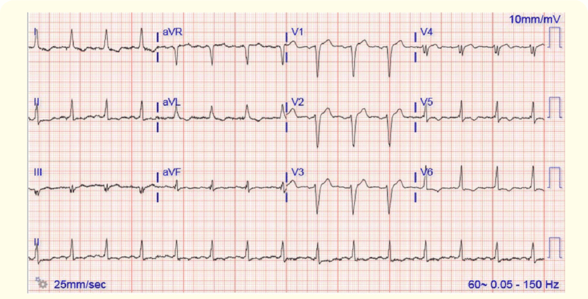
- Leads V1-V2: The QRS complex appears as the letter M. More specifically, the QRS complex displays rsr, rsR or rSR pattern . Occasionally the S-wave does not reach the baseline. The second R-wave is virtually always larger than the first R-wave.
- Leads V5, V6, I, aVL: Broad S-wave. S-wave duration is greater than R-wave duration, or S-wave duration is greater than 40 ms in V6 and I.
- ST-T changes: V1-V2 shows downsloping ST-segments and inverted T-waves. Leads V5, V6, I and aVL shows positive T-waves.
If the QRS duration is 0,110 seconds but < 0,12 seconds, the right bundle branch block is said to be incomplete. Note that a second r-wave may occur as a normal variant in lead V1 . Moreover, the normal septal q-waves are not affected by right bundle branch block. Occasionally the right bundle branch block only displays a broad and notched R-wave in V1 in that scenario the R-wave peak time should be > 0.05 seconds.
Is Bifascicular Block Dangerous
The main complication of bundle branch block, right or left, is to progress to a complete block of the electric conduction from the upper chambers of the heart to the lower, also called complete heart block or third-degree heart block. This can slow your heart rate, which can cause fainting and lead to serious complications and abnormal heart rhythms.
You May Like: How Does Sinus Pressure Make You Feel
Sinus Rhythm With Other Disturbances
When we speak about normal sinus rhythm, the adjective ânormalâ refers only to heart rhythm, it does not mean that the complete EKG is normal.
For example, normal sinus rhythm can be described in the presence of bundle branch block or signs of acute myocardial infarct.
It is a different matter when an initial sinus rhythm is associated with other heart rhythm disturbance as AV blocks, conduction via accessory pathway or pacemaker pacing.
In these conditions, the diagnosis of sinus rhythm should be restricted to atrial rhythm only.
In these cases is possible to determine sinus rhythm because P waves fulfill the first three criteria described above, replacing R-R interval with P-P interval.
- Heart rate between 60 and 100 bpm .
- P-P interval must be constant .
- Positive P wave in lead II and negative in lead aVR.
Is Right Bundle Branch Block Permanent

Yes.
A note from Cleveland Clinic
Right bundle branch block isnt a cause for worry if you dont have symptoms or heart disease. But when you have right bundle branch block and youve already had a heart attack or heart failure, you should focus on continuing with treatment for those problems. Be sure to follow your providers treatment plan for the best results.
Don’t Miss: What Is A Sinus Head Cold
What Is A Sinus Rhythm With A Right Bundle Branch Block
Ask U.S. doctors your own question and get educational, text answers â it’s anonymous and free!
Ask U.S. doctors your own question and get educational, text answers â it’s anonymous and free!
HealthTap doctors are based in the U.S., board certified, and available by text or video.
Right Bundle Branch Block
The right side of the heart receives deoxygenated blood from the body’s circulation into the right atrium and sends this blood to the right ventricle, and then to the lungs to be replenished with oxygen.
The contraction of the right ventricle is normally slightly less powerful than the contraction of the left ventricle. A right bundle branch block disrupts the contraction of the right ventricle.
Recommended Reading: Advil Sinus Congestion And Pain Walmart
Sinus Rhythm With Ventricular Arrhythmias
During a ventricular tachycardia or an accelerated idioventricular rhythm, a basal sinus rhythm can be seen with atrioventricular dissociation. P waves are usually difficult to distinguish..
The P wave rate is lower than ventricular rate.
The A-V dissociation in a broad QRS complex tachycardia is one of the diagnostic criteria of ventricular tachycardia.
References
- 1. Surawicz B, Knilans T. Chouâs electrocardiography in clinical practice. 7th ed. Philadelphia: Saunders Elservier 2008.
If you Like it… Share it.
The Heart’s Electrical System
The heart has four chambers that pump rhythmically by sequentially contracting and relaxing to circulate blood throughout the body and the lungs. Heart muscles are controlled by the cardiac electrical system, which is a branched distribution of nerves embedded in the heart muscle.
The sinus node is a bundle of nerves located in the right atrium. It controls the heart’s electrical system by sending signals across the heart’s left and right atria, stimulating them to contract. The message also passes through the atrioventricular node to the ventricles via a band of cardiac nerve fibers called the bundle of His.
The right and left bundle branches distribute the electrical impulse from the bundle of His across the right and left ventricles, causing them to beat. When the bundle branches are functioning normally, the right and left ventricles contract regularly and nearly simultaneously. This is described as normal sinus rhythm.
You May Like: How To Relieve A Sinus Migraine
Sinus Rhythm With Pre
Patients with accessory pathway have a double atrioventricular conduction: AV node and accessory pathway, this cause a short PR interval and a delta wave on the EKG .
It is not a normal sinus rhythm if signs of pre-excitation are seen on the electrocardiogram.
It may be described as sinus rhythm with short PR interval with delta wave.
Clinical Significance Of Right Bundle Branch Block
Right bundle branch block in asymptomatic individuals is not correlated with adverse outcomes. On the other hand, new right bundle branch block in patients with chest pain may indicate occlusion in the left anterior descending artery. Finally, new right bundle branch block in patients experiencing dyspnea may indicate pulmonary embolism. In the vast majority of cases, however, right bundle branch block is a benign finding with little if any impact of cardiovascular prognosis. Young individuals rarely display right bundle branch block. However, incomplete right bundle branch block occurs occasionally even in younger persons. A large prospective cohort study evaluated the association between right bundle branch block and mortality over a period of 20 years in otherwise healthy individuals no association was found.
Don’t Miss: The Best Over The Counter Sinus Medication
Sinus Rhythm With Ventricular Pacemaker Pacing
The patient with electrical pacemaker may have sinus rhythm in the atria with ventricular pacing.
In dual-chamber pacemakers programmed in DDD mode ventricular pacing must follow the sinus P wave.
In single-chamber pacemaker , if there were sinus activity, ventricular pacing would be independent of atrial rhythm.
In both cases we may describe the EKG as sinus rhythm with ventricular pacing by electrical pacemaker.
In patients with pacemaker, if pacing is inhibited by sensed spontaneous cardiac depolarizations, we can describe normal sinus rhythm.
B26 Left Bundle Branch Block

The ECG was recorded from a 35 year old man who had presented with a six month history of chest pain and lightheadedness on exertion
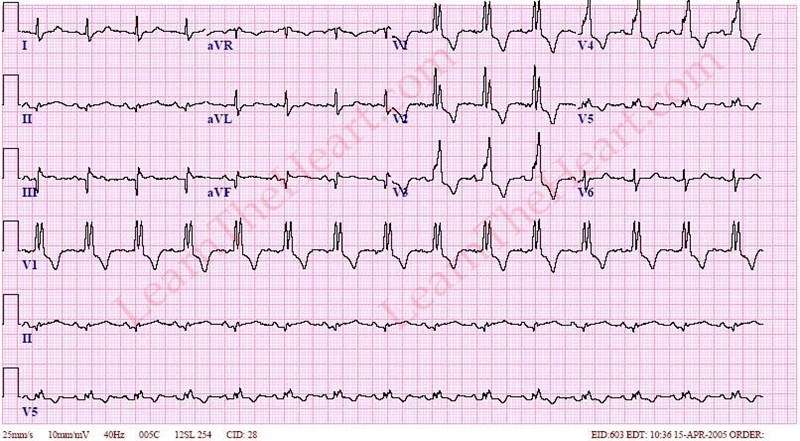
ECG. Rhythm: Sinus rhythm is present, all beats are conducted with a normal PR interval. The ninth complex in the rhythm strip occurs earlier than expected. The QRS complex is of the usual configuration for this patient but the P wave that precedes it is different to the others in this lead. An ectopic focus within the atria is responsible for initiating atrial contraction and gives rise to an atrial premature beat.
Morphology: The QRS duration is prolonged beyond the upper limit of normal . The normal Q wave in lead V5 or V6 that represents septal depolarization is absent. There is no secondary R wave in VI as occurs in RBBE. These are the diagnostic features of left bundle branch block. Several other ECG changes that may occur in LBBB are seen in this ECG although they are not essential to make the diagnosis. The QRS complexes in leads facing the left ventricle show an M shaped pattern and there are secondary changes in the ST segments which are depressed and accompanied by T wave inversion. The initial R waves that are normally seen in the right sided precordial leads are absent in this record and the complexes in these leads are changes in these leads the ST segments are elevated and the T waves are tall. As explained elsewhere these changes should not be interpreted to indicate ischaemia in the presence of LBBB.
Also Check: Young Living Oils For Sinus Pressure
Normal Sinus Rhythm With Left Bundle Branch Block
77 year old male patient monitored during colectomy. Patient has a history of coronary artery disease.
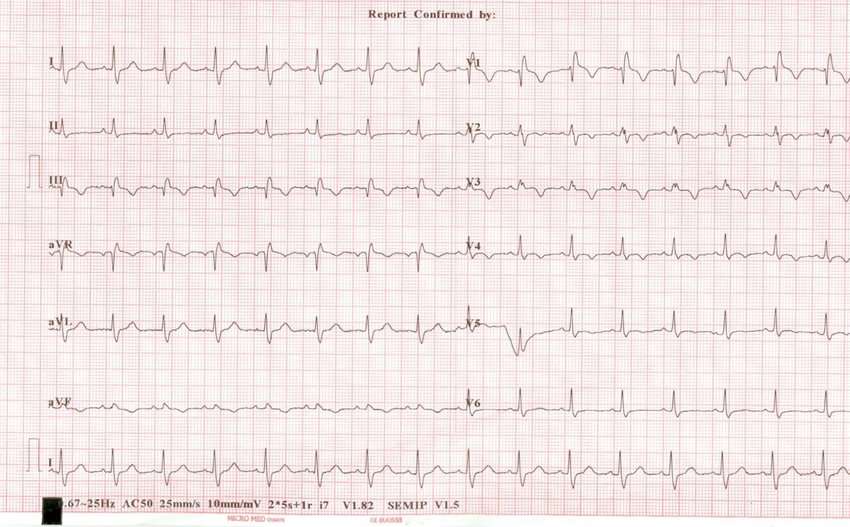
Rhythm analysis indicates a normal sinus rhythm . First degree heart block is present, as well as a left bundle branch block.
This encounter shows normal sinus rhythm with P waves and a regular rhythm. The PR interval is greater than 200 milliseconds, making this first degree heart block. Additionally, the QRS is wider than 120 milliseconds, leading to a further investigation of heart blocks. The QRS to the left of the J point in lead V1 is negative, making this a left bundle branch block.
EKGmon is a telemetry monitoring and quiz platform. It simulates EKG monitors found in hospitals, by streaming EKG data to a display in real time.
Both information and quiz modes are available from the top menu. Speed and amplitude of the waveforms can be adjusted to better view telemetry data.
EKGmon has several distinct advantages over traditional EKG training methodologies. The data provided by EKGmon only comes from real patient encounters. No data is simulated.
The EKG data here is presented in a streaming fashion, very similar to hospital telemetry monitors. This is far different than other methodologies which only utilize static 6-second strips for training.
Dual signals are also provided, typically on Leads II and V1. This allows for an immersive experience most similar to that of a hospital EKG tech or nurse.
| Name |
|---|
What Are The Symptoms
Left bundle branch block often doesnt cause any symptoms. In fact, some people have it for years and never know they have the condition.
For others, however, a delay in the arrival of electrical impulses to the hearts left ventricle can cause syncope , due to unusual heart rhythms that affect blood pressure.
Some people might also experience something called presyncope. This involves feeling like youre about to faint, but never actually fainting.
Other symptoms can include fatigue and shortness of breath.
Recommended Reading: Advil Sinus And Pain Dosage
What Is The Outlook For Right Bundle Branch Block
Your outlook depends on whether you have cardiovascular disease. If you dont have heart disease, having right bundle branch block doesnt change your life expectancy or add to your risk level. But having right bundle branch block can put you at a higher risk of death if you also have heart failure or a heart attack.
What Causes Right Bundle Branch Block
Right bundle branch block can result from a number of conditions, such as:
-
Heart disease from high blood pressure
-
Chronic obstructive lung disease
-
Pulmonary embolism
-
Congenital heart disease
-
Surgery or other procedures on the heart
In some case, the cause of right bundle branch block is not known. The heart otherwise appears normal.
You May Like: Sinus Pressure Relief For Kids
Rbbb And Acute Myocardial Infarction
The RBBB pattern occurs in 3 to 29 percent of patients with acute myocardial infarction17 . It is often accompanied by left anterior fascicular block.18 The important lesion is usually in the left anterior descending coronary artery.19 A mortality rate of 36 to 61 percent has been reported in these patients. Hindman and associates reported an in-hospital mortality of 24 percent and a total 1-year mortality of 48 percent in patients with acute myocardial infarction and RBBB.20 The time of onset of the RBBB in relation to infarction was uncertain in many cases. Several studies have shown that the mortality rate was higher in patients with new-onset RBBB than in those with an old RBBB. This is in contrast to the findings in patients with acute anterior myocardial infarction and LBBB, for whom the mortality rate with an old LBBB was higher than in those with recent-onset bundle branch block.19
Brian Olshansky MD, … Nora Goldschlager MD, in, 2017
How Is Left Bundle Branch Block Treated
Left bundle branch block doesnt always require treatment, especially if you dont have any underlying heart conditions.
If you do have another heart condition, your doctor might suggest treating the underlying cause or no treatment at all if youre stable.
If you have left bundle branch block due to electrical problems with your conduction system, for example, you may need a pacemaker. This is a device that emits electricity to help your heart maintain a consistent rhythm.
If you have high blood pressure, you may need to take medication to keep it under control. This will also help reduce the strain on your heart.
While treating the underlying condition might not completely get rid of left bundle branch block, it can lessen the risk of complications, such as progressive disease.
Don’t Miss: Sinus Or Cold Or Allergy
Pr Interval Higher Than Or Equal To 012 S
Short PR intervals with delta waves are signs of the existence of an accessory pathway .
It is not considered normal sinus Rhythm because the stimulus is conducted by a structure outsider the normal conduction system.
There may also be short PR interval in low atrial rhythm or in retrograde atrial conduction due to a junctional rhythm, but neither is sinus rhythm.
Normal Sinus Rhythm and Long PR interval:
There is no general agreement on whether the long PR interval rules out normal sinus rhythm.
The majority of authors accept it as Normal Sinus Rhythm, because the stimulus is conducted by normal conduction system.
It may be described as sinus rhythm with first degree AV block or long PR interval.
Living With Right Bundle Branch Block

Your doctor may give you more instructions about how to manage your overall heart health. You might need to make lifestyle changes. These may include losing weight, quitting smoking, or improving your diet.
Keep track of any heart symptoms you have. See your doctor regularly, even if you dont have any symptoms. Make sure all your healthcare providers know about your right bundle branch block.
You May Like: Where Does Sinus Cancer Spread To
Clinical Implications Of Left Bundle Branch Block
Left bundle branch block is always pathological. It affects left ventricular contractility and pumping function. Consequently, left bundle branch block confers adverse cardiovascular outcomes. Left bundle branch block is associated with hypertension, ventricular hypertrophy, valvular heart disease, myocarditis, ischemic heart disease, heart failure and cardiomyopathies. The Framingham Heart Study showed that acquired left bundle branch block was associated with seven times as great a risk of heart failure, two times as great a risk of coronary artery disease and significantly higher risk of developing right ventricular hypertrophy. Left bundle branch block is rare in young individuals and appears to affect their prognosis little.
Bifascicular Blocks What You Need To Know
Anatomy of the Hearts Electrical Conduction System
Ventricular depolarizaiton is facilitated by the hearts electrical conduction system, sometimes referred to as the His/Purkinje system, which by convention is said to have three main fascicles or branches. There is a single fascicle in the right ventricle referred to as the right bundle branch. There are two fascicles in the left ventricle: the left anterior fascicle and the left posterior fascicle of the left bundle branch.
It reality, while it is clinically useful to think in terms of a left anterior fascicle and a left posterior fascicle, anatomically speaking the left bundle branch has many fascicles that fan into the the left ventricle as shown by these drawings by Sunao Tawara, the Japanese physician who discovered the AV node.
Lets start by looking at what a single fascicle looks like when it is blocked. This is sometimes referred to as a hemiblock, or a hemifascicular block. To identify fascicular blocks you need to be able to identify a right or left axis deviation in the frontal plane. One way to do that is to use a cheat sheet like this one.
Left Anterior Fascicular Block
Rules for left anterior fascicular block:
- Modest widening of the QRS complex
- Left axis deviation
- rS complexes in the inferior leads
- qR complexes in the high lateral leads
Left Posterior Fascicular Block
Rules for left posterior fascicular block:
However, isolated left posterior fascicular block is very rare.
Right Bundle Branch Block
You May Like: Can I Treat A Sinus Infection Without Antibiotics