What Are The Signs And Symptoms Of Skull Base Tumor
Symptoms appear slowly as the tumor grows and puts pressure on vital structures in the brain such as the pituitary gland, the optic nerve and the carotid arteries.
Specific symptoms depend on the type, location and size of the tumor. For example, tumors involving the skull base and nose can affect breathing and sense of smell. Some tumors in the pituitary gland can affect vision and swallowing.
In general, common symptoms of skull base tumors include:
How Is The Procedure Performed
The technologist begins by positioning patients on the CT examination table.
For a CT scan of the sinuses, the patient is most commonly positioned lying flat on the back. The patient may also be positioned face-down with the chin elevated.
Straps and pillows may be used to help the patient maintain the correct position and to hold still during the exam.
Some patients require an injection of a contrast material to enhance the visibility of certain tissues or blood vessels. If contrast material is required, a nurse or technologist will insert an intravenous line into a small vein in the patient’s hand or arm. The contrast material will be injected through this line.
Next, the table will move quickly through the scanner to determine the correct starting position for the scans. Then, the table will move slowly through the machine for the actual CT scan. Depending on the type of CT scan, the machine may make several passes.
The technologist may ask you to hold your breath during the scanning. Any motion, including breathing and body movements, can lead to artifacts on the images. This loss of image quality can resemble the blurring seen on a photograph taken of a moving object.
When the exam is complete, the technologist will ask you to wait until they verify that the images are of high enough quality for accurate interpretation by the radiologist.
The actual CT scan takes less than a minute and the entire process is usually completed within 10 minutes.
Can A Ct Scan Of Sinuses Show A Brain Tumor
This is useful if sinusitis is suspected. A typical series of CT scans for the sinuses use less x-ray radiation than a standard complete set of x-rays. However, a CT scan of the sinuses does not show any brain tissue.
What is the most common CT scan?
The following CT scans are either sometimes or always done with IV and/or oral contrast and will require preparation: CT Abdomen most commonly used to diagnose and monitor cancer, abdominal pain, and bowel obstruction. CT Pelvis most commonly used to diagnose and monitor cancer, pelvic pain, and bowel obstruction.
Recommended Reading: What Can I Take For Sinus Pressure
Your Complete Ct Scan Experience
Upon your arrival, your technician will take you to a private treatment room where you will sit upright in a chair.
Your technician will place a lead apron onto your body to protect you. They will ask you to remove any earrings, jewelry, and/or hearing aids. Your head will then be positioned inside the scanner.
The MiniCAT scanner is an open device, so theres no risk of claustrophobia, which is common during traditional CT scans. Once the machine is turned on, the scan takes just 20 seconds and uses the lowest radiation dose possible. Your entire appointment will last about five minutes, at which point you will be free to return to work and resume your regular daily routine.
Incisional And Excisional Biopsies

These types of biopsies remove more of the tumor using minor surgery. They’re the more common types of biopsies done for nasal and paranasal sinus tumors. Biopsies of tumors in the nose may be done using special tools that are put into the nose. Biopsies of tumors that are deeper within the skull may require a more involved procedure .
For an incisional biopsy, the surgeon cuts out a small piece of the tumor. For an excisional biopsy, the entire tumor is removed. In either case, the biopsy sample is then sent to the lab for testing.
Also Check: Antibiotics Not Helping Sinus Infection
Positron Emission Tomography Scan
A PET scan uses a slightly radioactive form of sugar that’s injected into your blood and collects mainly in cancer cells. A special scanner is then used to create pictures of the places where the radioactivity collected in your body.
A PET scan may be used to look for possible areas of cancer spread, or if a CT or MRI scan does not show an obvious tumor. This test also can be used to help see if a change seen on another imaging test is cancer or not.
PET/CT scan: A PET scan is often done along with a CT scan using a machine that can do both scans at the same time. This lets the doctor compare areas of higher radioactivity on the PET scan with the detailed pictures from the CT scan.
What Will I Experience During And After The Procedure
CT exams are generally painless, fast, and easy. Multidetector CT reduces the amount of time that the patient needs to lie still.
Though the scanning itself causes no pain, there may be some discomfort from having to remain still for several minutes. If you have a hard time staying still, are claustrophobic, or have chronic pain, you may find a CT exam to be stressful. The technologist or nurse, under the direction of a physician, may offer you some medication to help you tolerate the CT scanning procedure.
If the exam uses iodinated contrast material, your doctor will screen you for chronic or acute kidney disease. The doctor may administer contrast material intravenously , so you will feel a pin prick when the nurse inserts the needle into your vein. You may feel warm or flushed as the contrast is injected. You also may have a metallic taste in your mouth. This will pass. You may feel a need to urinate. However, these are only side effects of the contrast injection, and they subside quickly.
When you enter the CT scanner, you may see special light lines projected onto your body. These lines help ensure that you are in the correct position on the exam table. With modern CT scanners, you may hear slight buzzing, clicking and whirring sounds. These occur as the CT scanner’s internal parts, not usually visible to you, revolve around you during the imaging process.
You May Like: Medicine For Runny Nose And Sinus Pressure
What Is A Ct Scan Of The Brain
A CT of the brain is a noninvasive diagnostic imaging procedure that usesspecialX-raysmeasurements to produce horizontal, or axial, images of the brain. Brain CT scans can provide more detailed information aboutbrain tissue and brain structures than standard X-rays of the head, thusproviding more data related to injuries and/or diseases of the brain.
During a brain CT, the X-ray beam moves in a circle around the body,allowing many different views of the brain. The X-ray information is sentto a computer that interprets the X-ray data and displays it in atwo-dimensional form on a monitor.
Brain CT scans may be done with or without “contrast.” Contrast refers to asubstance taken by mouth or injected into an intravenous line thatcauses the particular organ or tissue under study to be seen more clearly.Contrast examinations may require you to fast for a certain period of timebefore the procedure. Your physician will notify you of this prior to theprocedure.
Other related procedures that may be used to diagnose brain disordersincludeX-rays,magnetic resonance imaging of the brain,positron emission tomography scan of the brain, andcerebral arteriogram.
Magnetic Resonance Imaging Scan
Like CT scans, MRI scans show detailed images of the body. But MRI scans use radio waves and strong magnets instead of x-rays. MRI scans are very helpful in looking at cancers of the nasal cavities and paranasal sinuses. They are better than CT scans in telling whether a change is fluid or a tumor. Sometimes they can help the doctor tell the difference between a lump that is cancer and one that is not. They can also show if a tumor has spread into nearby soft tissues, like the eyeball, brain, or blood vessels.
Read Also: Is It A Toothache Or Sinus Infection
Outlook For Nasal And Sinus Cancer
There are many different types of cancer that can affect the nasal cavity and sinuses.
The outlook varies, depending on the specific type of nasal and sinus cancer you have, its exact location, how far it’s spread before being diagnosed and treated, and your overall level of health and fitness.
The Cancer Research UK website has more information about the outlook for nasal and sinus cancer.
Nasal And Sinus Cancer
Nasal and sinus cancer is a rare cancer that affects the nasal cavity and the sinuses .
Nasal and sinus cancer is different from cancer of the area where the nose and throat connect.
This is called nasopharyngeal cancer.
Gwen Shockey/SCIENCE PHOTO LIBRARY https://www.sciencephoto.com/media/704091/view
You May Like: Advil Sinus Congestion And Pain Drowsiness
Mucous Retention Cysts Mucoceles And Nasal Polyps
Different kinds of masses can be found inside the nose and sinus. While some might be benign and not lead to complications, others might be the reason forrecurrent sinus problems.Mucous retention cystsare fluid-filled benign cysts, often found in the maxillary sinus.
Mucocelesare thick liquid-filled walled-off sinus cells that can lead to complications such as compression of the eye and sinus headaches. I usually treat mucoceles with expandedendoscopic sinus surgerytechniques that limit complications and decrease the risk of recurrence.
Nasal polypsare noncancerous growths that look like water-filled growths and generally do not cause pain. They result from sinus inflammation and could be the main reason for recurring sinus problems.Nasal polyps require expert management, often utilizing both surgical and medical therapies.
It is necessary to identify each type of cyst to prevent them from leading to further complications.
Treatments For Nasal And Sinus Cancer

The treatment recommended for you will depend on several factors, including the stage at which the cancer was diagnosed, how far it’s spread, and your general level of health.
Treatment may include:
- surgery to remove a tumour this can be performed through open surgery or as keyhole surgery through the nose
- radiotherapy where high-energy radiation is used to kill the cancerous cells, shrink a tumour before surgery, or destroy small pieces of a tumour that may be left after surgery
- chemotherapy where medicine is used to help shrink or slow down the growth of a tumour, or reduce the risk of the cancer returning after surgery
If you smoke, it’s important that you give up.
Smoking increases your risk of cancer returning and may cause you to have more side effects from treatment.
Your treatment will be organised by a head and neck cancer multidisciplinary team , who’ll discuss the treatment options with you.
A combination of treatments will often be recommended.
Read Also: What Is The Best Pain Reliever For Sinus Pressure
Mucosal Thickening And Opacification
Mucosal thickening is an inflammatory reaction in your sinuses. It can be triggered by allergic reactions, infection, ornasal polyps. It may also lead to opacification in your sinuses and cause further complications. Therefore, a sinus CT scan is necessary to diagnose mucous thickening and opacification.
How Does The Procedure Work
In many ways, a CT scan works like other x-ray exams. Different body parts absorb x-rays in different amounts. This difference allows the doctor to distinguish body parts from one another on an x-ray or CT image.
A conventional x-ray exam directs a small amount of radiation through the body part under examination. A special electronic image recording plate captures the image. Bones appear white on the x-ray. Soft tissue, such as the heart or liver, shows up in shades of gray. Air appears black.
With CT scanning, several x-ray beams and electronic x-ray detectors rotate around you. These measure the amount of radiation being absorbed throughout your body. Sometimes, the exam table will move during the scan. A special computer program processes this large volume of data to create two-dimensional cross-sectional images of your body. The system displays the images on a computer monitor. CT imaging is sometimes compared to looking into a loaf of bread by cutting the loaf into thin slices. When the computer software reassembles the image slices, the result is a very detailed multidimensional view of the body’s interior.
Nearly all CT scanners can obtain multiple slices in a single rotation. These multi-slice CT scanners obtain thinner slices in less time. This results in more detail.
For children, the radiologist will adjust the CT scanner technique to their size and the area of interest to reduce the radiation dose.
Don’t Miss: Advil Cold And Sinus 200 Mg
What Are The Risks Of A Ct Scan Of The Brain
You may want to ask your doctor about the amount of radiation usedduring the brain CT procedure and the risks related to your particularsituation. You should keep a record of your past history of radiationexposure, such as previous CT scans and other types of X-rays, so thatyou can inform your doctor. Risks associated with radiation exposuremay be related to the cumulative number of X-ray examinations and/ortreatments over a long period of time.
To safeguard your health, consider the following precautions beforescheduling a brain CT:
There may be other risks depending on your specific medical condition.Be sure to discuss any concerns with your doctor prior to theprocedure.
How Should I Prepare
Wear comfortable, loose-fitting clothing to your exam. You may need to change into a gown for the procedure.
Metal objects, including jewelry, eyeglasses, dentures, and hairpins, may affect the CT images. Leave them at home or remove them prior to your exam. Some CT exams will require you to remove hearing aids and removable dental work. Women will need to remove bras containing metal underwire. You may need to remove any piercings, if possible.
Your doctor may instruct you to not eat or drink anything for a few hours before your exam if it will use contrast material. Tell your doctor about all medications you are taking and if you have any allergies. If you have a known allergy to contrast material, your doctor may prescribe medications to reduce the risk of an allergic reaction. To avoid unnecessary delays, contact your doctor well before the date of your exam.
Also tell your doctor about any recent illnesses or other medical conditions and whether you have a history of heart disease, asthma, diabetes, kidney disease, or thyroid problems. Any of these conditions may increase the risk of an adverse effect.
Women should always inform their physician and the CT technologist if there is any possibility that they may be pregnant. See the CT Safety During Pregnancy page for more information.
Recommended Reading: Treatment For Sinus Infection In Adults
Medical History And Physical Exam
You will be asked about your medical history, any problems you’ve been having, and possible risk factors such as where you work and what chemicals you work with. The doctor will physically examine you to look for signs of nasal cavity or paranasal sinus cancer, as well as other health problems.
During the exam, the doctor will carefully check your head and neck area, including the nose and sinuses, for numbness, pain, swelling, and/or firmness in your face and the lymph nodes in your neck. The doctor will look for changes in the symmetry of your eyes and face , vision changes, and any other problems.
What Is The Cost Of Ct Pns In India
The cost of CT PNS ranges from 1000 to 7000 INR on average. However, the variation in cost depends upon the city and the laboratory where the procedure is done.
What is a CTCT scan of the nervous system?
CT Scan PNS Axial and Coronal with Contrast Test. The peripheral nervous system and central nervous system are the two components of the nervous system .The main function of the PNS is to connect the CNS to the limbs and organs, acting as serving bridge between the brain and spinal cord and the rest of the body.
You May Like: What To Take For Sinus Infection While Pregnant
Diagnosing Nasal And Sinus Cancer
Tests you may have to help diagnose nasal and sinus cancer include:
- a nasal endoscopy where a long, thin, flexible tube with a camera and light at the end is inserted into your nose to examine the area this can be uncomfortable, so before the procedure you’ll be asked whether you’d like anaesthetic sprayed on the back of your throat
- a biopsy where a small sample of tissue is removed and examined this may be done during an endoscopy
- a fine needle aspiration where fluid and cells are taken from a lymph node using a needle to see if the cancer has spread
If you’re diagnosed with nasal and sinus cancer, you may have a CT scan, MRI scan, PET scan or ultrasound scan to help stage and grade the cancer.
What Are The Different Types Of Skull Base Tumor

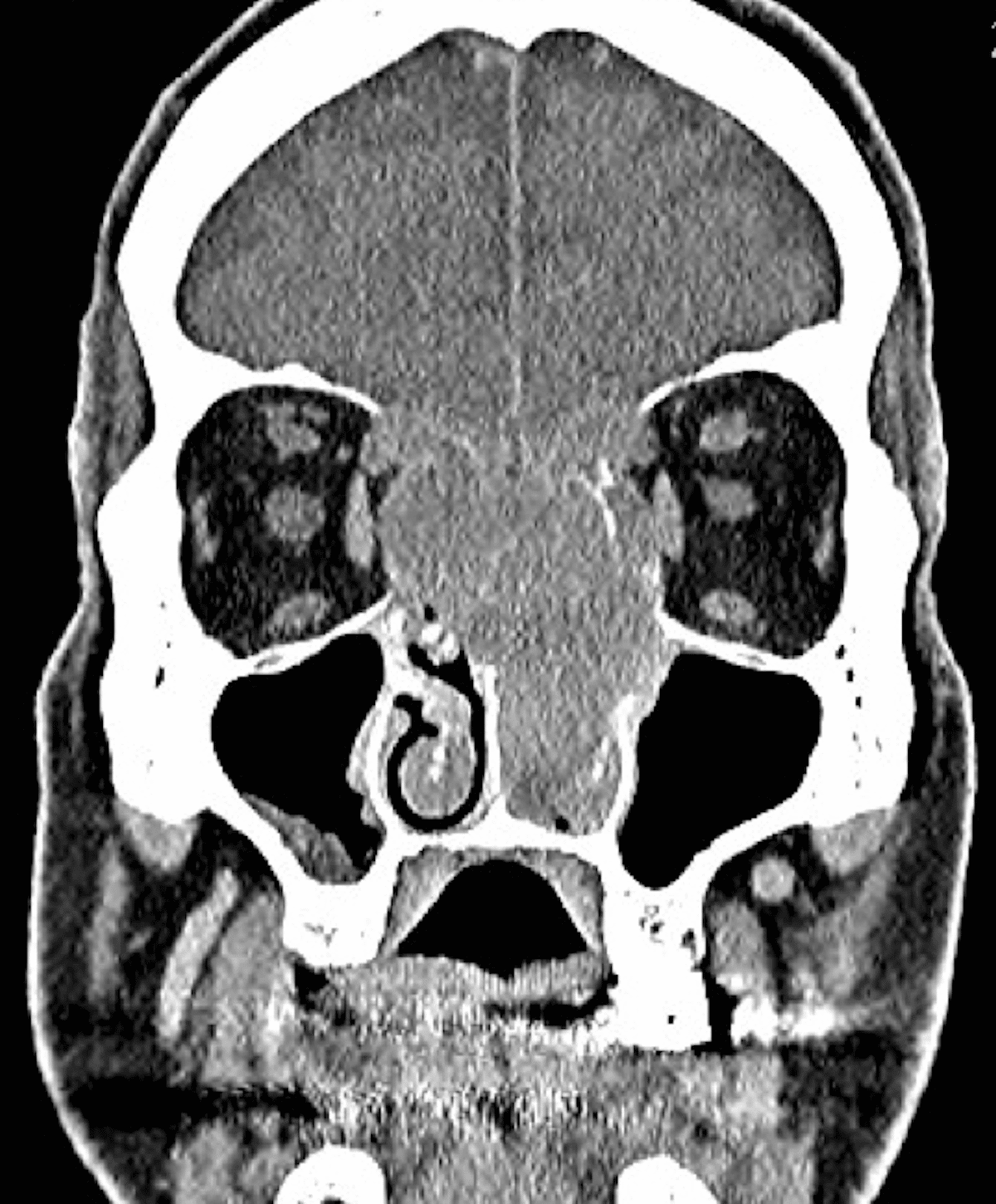
Skull base tumors most often grow inside the skull but occasionally form on the outside. They can originate in the skull base as a primary tumor or spread there from a cancer elsewhere in the body as a metastatic brain tumor.
Skull base tumors are classified by tumor type and location within the skull base.
In the front section of the skull base , which contains the eye sockets and sinuses, the following tumors are more likely:
Read Also: What Is Good To Take For Sinus Headache
What Is A Computed Tomography Scan Of The Pns
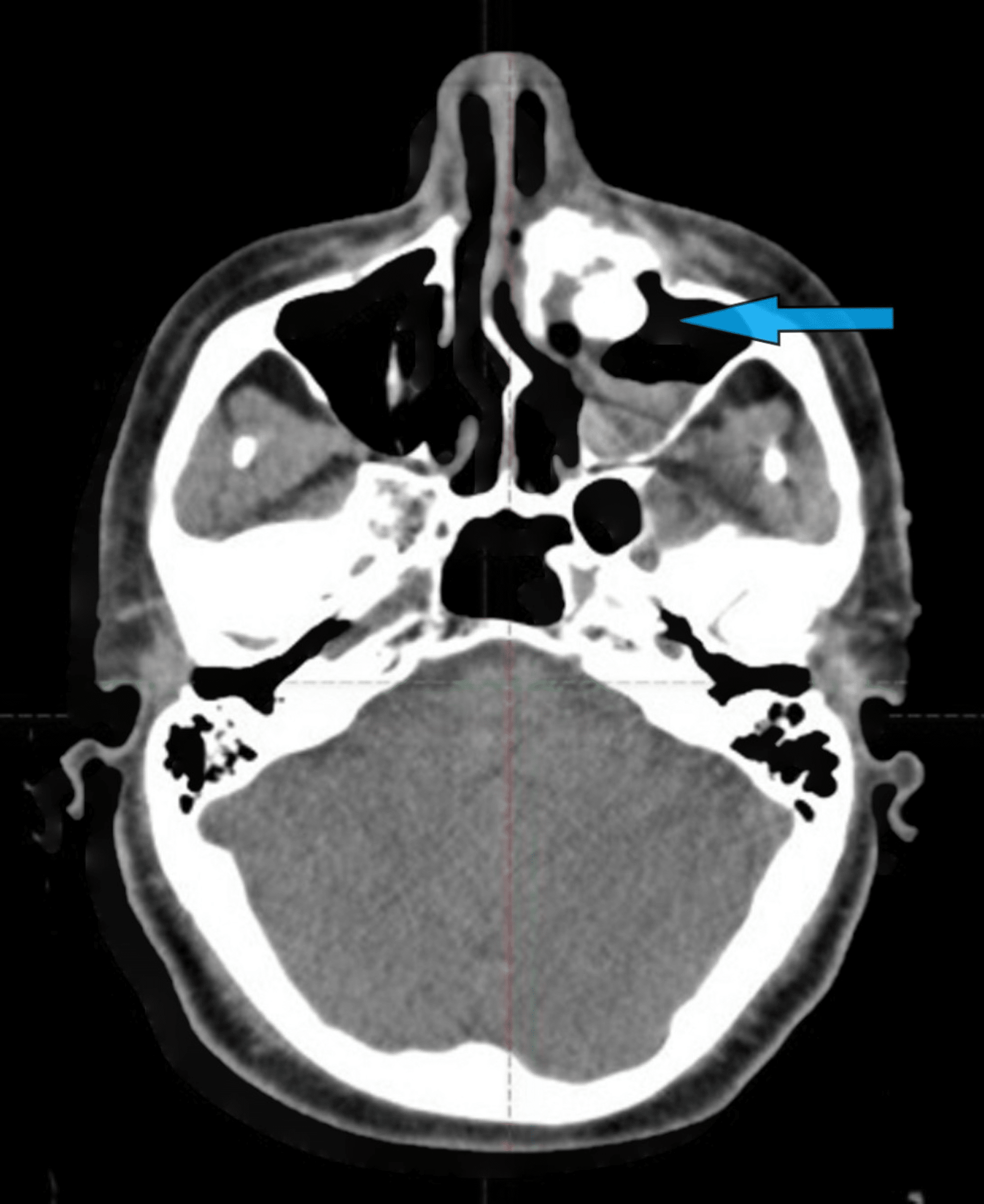
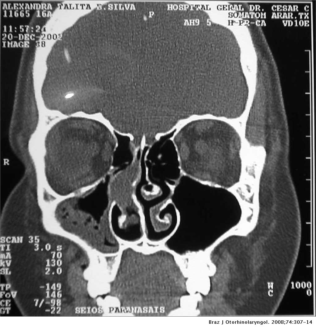
A Computed Tomography Scan of the PNS is an imaging test of sinuses which uses X-Rays to bring out in-depth images of air-filled spaces within the bones of the face, surrounding the nasal cavity. It usually includes the upper area of the throat, behind the nose.
What is CTCT PNS and how is it used?
CT PNS is the most commonly used imaging technique in the field of otolaryngology. The test has been into use to detect and study the abnormalities in paranasal sinuses.